What is “Homology”
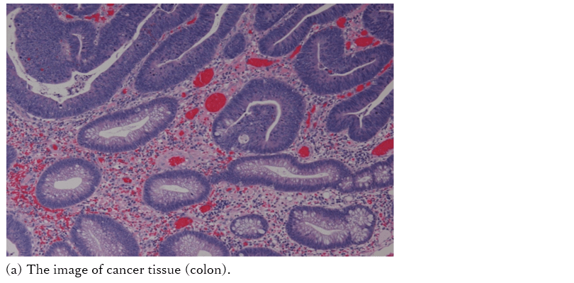
Homology is a mathematical concept that starts with the problem of the “Königsberg Bridge“. The basic principle is “the number of enclosed areas” implies “the degree of contact”. Let’s use figures (a), (b) and (c) to explain the concept.

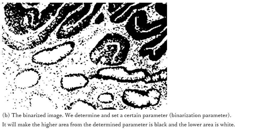
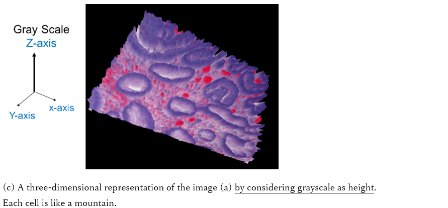
If we denote the the number of isolated regions by b0 and the number of enclosed areas by b1, figures (a), (b) and (c) could be described as follows.
(a) b0=7, b1=0 and b1/b0=0: The regions are separated from one another.
(b) b0=3, b1=1 and b1/b0=0.333: Several regions contact one another.
(c) b0=1, b0=2、b1/b0=2.0: If regions contact further more.
In other words, the degree of contact can be quantitatively evaluated by b1 and the ratio b1/b0 (this ratio gives number of surrounded areas in one lump). It is important that this method does not consider the shape of the “surrounded areas”. This property is called “the topological invariant”. Because of this property, we can apply this method on images having various forms such as cancer tissues.
The relation of cancer and the homology
Cancer has the characteristic property of loss of contact inhibition. This means that cancer cells do not stop growing on contact (read more).The variety of forms of cancer tissue is due to this property. By using the homology value b0 and b1, we detect the areas where the contact degree is high.
The following is a schematic explanation how to measure the contact of cells. In the bottom of the figure, the area above certain contour lines are blacked out.
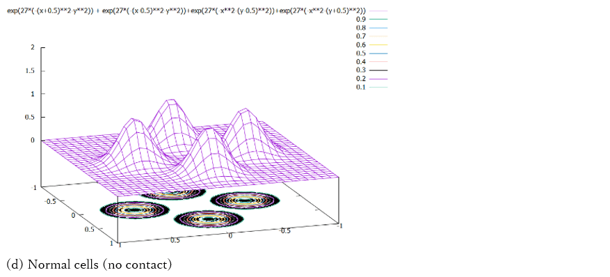
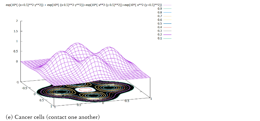
Since the mountain parts are separated from one another in figure (d), there is no surrounded area at any height (binarized parameter). If they are very close like in figure (e), at a certain height (binarized parameter), there is a surrounded area. This situation occurs when cancer cells overlap or come into contact.
Advantage of using Homology Profile
- Robust algorithm
- Low computation cost and fast runtime
- “White box” algorithm
- No need to collect huge number of annotated training data for algorithm development
Research Publications
- Homology analysis detects topological changes of Iba1 localization accompanied by microglial activation Sawano T, Tsuchihashi R, Morii E, Watanabe F, Nakane K, Inagaki S, Neuroscience volume 346, issue, pp. 43 – 51 2017.
- Homology-based method for detecting regions of interest in colonic digital images K.Nakane, A.Takiyama,S Mori, N.Matuura, Diagnostic Pathology 2015, 10:36 2015.
- Fast and Accurate Tumor Segmentation of Histology Images using Persistent Homology and Deep Convolutional Features, Qaiser T, Taniyama D, Epstein D, Nakane K, Sakamoto K, Rajpoot N, Yee-Wah Tsang, Medical Image Analysis, Volume 55, Pages 1-14 2019.
- Automated gleason grading on prostate biopsy slides by statistical representations of homology profile, Yan C, Nakane K, Wang X, et al. Comput Methods Programs Biomed. 2020;194:105528.
- Automated prediction of emphysema visual score using homology-based quantification of low-attenuation lung region, Nishio, Mizuho et al. PloS one vol. 12,5 e0178217. 25 May. 2017, doi:10.1371/journal.pone.0178217
- Homology-based radiomic features for prediction of the prognosis of lung cancer based on CT-based radiomics, Kadoya N, Tanaka S, Kajikawa T, et al. Med Phys. 2020; 47(5):2197-2205.2.
APSAM Application Areas
- Histology
- Colon cancer
- Prostate cancer (Gleason Score)
- Lymph node metastasis
- Lung cancer (Cytology, CT-image)